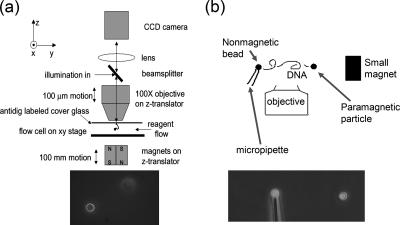
Figure 2.
MT setups. (a) Vertical MT system. A DNA molecule is suspended from the top surface of a flow cell by one end, with its other end attached to a 3-μm-diameter paramagnetic particle that acts as a “handle” to which controlled forces can be applied, by using a permanent magnet below the flow cell. The bead is observed using a microscope objective; a piezoelectric focuser is used to locate the bead in the vertical (z) direction. Illumination of the bead is done through the objective and therefore a reflected-light (dark-field) image is obtained (bottom). Buffer exchanges are carried out using flow through the sample cell. (b) Transverse MT system. The principle is the same as for the vertical tweezer, but with the modifications that in addition to the paramagnetic particle at the right end of the molecule, a nonmagnetic particle is attached at its left end, held using suction on a thin glass capillary. By using a small magnet that can be positioned in the same plane or even below the end of the pipette, forces on the paramagnetic bead can be applied with direction in the plane of focus. The result is that both ends of the DNA molecule can be imaged simultaneously (bottom). An open sample cell is used allowing straightforward removal and replacement of buffer.

