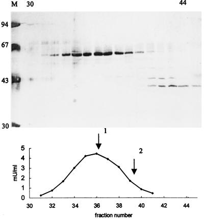
Figure 3.
Purification of 2-HPCL. The lyase was purified from a peroxisomal matrix protein fraction by successive chromatographic steps as described in Methods. (Upper) The polypeptide pattern of fractions 30–45 from the final gel filtration column as analyzed by SDS/PAGE (100 μl fraction; 10–20% gradient gel; silver staining). The migration of molecular mass (expressed in kDa) markers is represented in lane M. (Lower) The elution of lyase activity, expressed in mU/ml, from the same gel filtration column. Elution of calibration proteins is indicated by arrows: 1, catalase (240 kDa); 2, lactate dehydrogenase (140 kDa).

