
Figure 2. All four mammalian arrestins bind Ca2+/CaM in cells.
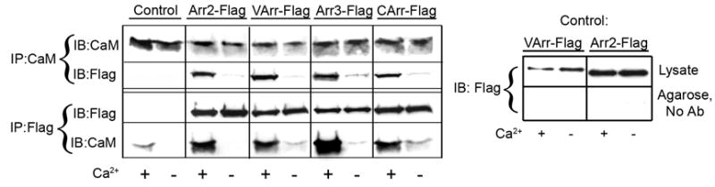
The lysates of cells expressing HA-CaM and the indicated flag-tagged arrestins (or empty vector; control) were immunoprecipitated (IP) with rabbit anti-Flag or rabbit anti-calmodulin (CaM) antibodies as described in the Methods. Aliquots of the total lysate and immunoprecipitated proteins were run on 12.5% (calmodulin) or 10% (arrestin) SDS-PAGE and blotted (IB) for HA-CaM or Flag-arrestin. Non-specific binding of arrestins to Protein G agarose (in the absence of antibody (No Ab)), was very low and calcium-independent (right panel). Visual (VArr), arrestin2 (Arr2), arrestin3 (Arr3), and cone arrestin (CArr) were expressed at 96+30, 74+26, 53+9, and 73+21 pmol/mg protein, respectively, yielding intracellular concentrations that are somewhat higher than those found in mature neurons for non-visual arrestins 39 and lower than those found in rod photoreceptors for visual arrestin 45; 54. HA-CaM was expressed at a level equal to that of endogenous CaM and was used to facilitate immunoblotting. Representative results from 2–4 experiments are shown.