
Figure 4. Summary of the changes in spin label mobility induced by arrestin interaction with Ca2+/CaM.
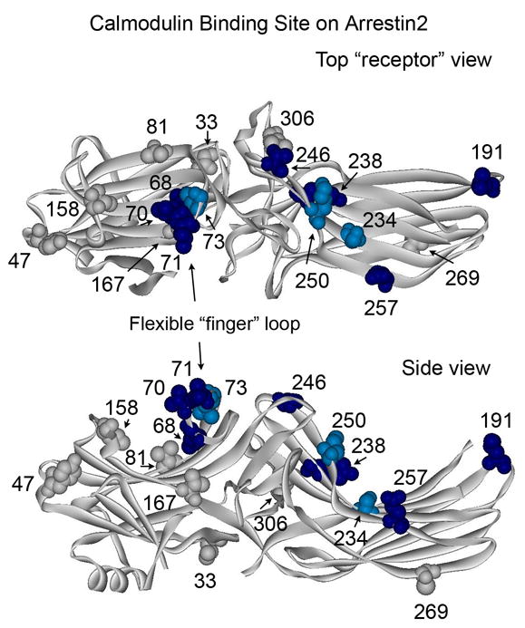
The magnitude of Ca2+/CaM-induced changes in spin label mobility (Fig.3) is color-coded on the arrestin2 crystal structure (PDB ID: 1G4R) 27 as follows: light blue/dark blue, small and large changes in mobility, respectively; gray, no change (spectral data not shown for sites 33, 47, 81, 167, 269, and 306). Top panel: view from the “receptor binding” side of arrestin. Bottom panel: side view.