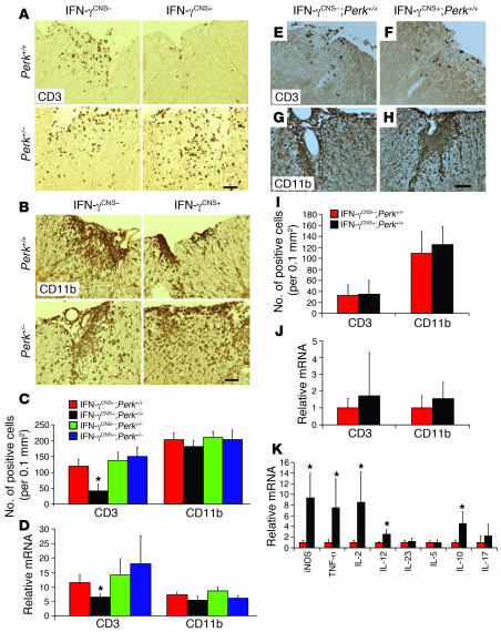
Figure 3. The effects of IFN-γ on inflammatory infiltration.
(A and C) CD3 immunostaining showed that CNS delivery of IFN-γ reduced T cell infiltration in the lumbar spinal cords of mice on a Perk+/+ background at PID17, but did not significantly affect T cell infiltration in mice on a Perk+/– background. (B and C) CD11b immunostaining revealed that CNS delivery of IFN-γ did not significantly change the numbers of CD11b-positive microglia/macrophages in the lumbar spinal cord of mice on a Perk+/+ or Perk+/– background at PID17 (n = 3). (D) Real-time PCR analysis of the relative mRNA levels of CD3 and CD11b in the spinal cord at PID17 (n = 3). (E, F, and I) CD3 immunostaining showed that CNS delivery of IFN-γ did not affect T cell infiltration in lumbar spinal cord at PID14. (G, H, and I) CD11b immunostaining showed that CNS delivery of IFN-γ did not significantly change the numbers of CD11b-positive microglia/macrophages in the lumbar spinal cord at PID14 (n = 3). (J) Real-time PCR analysis of the relative mRNA levels of CD3 and CD11b in the spinal cord at PID14 (n = 4). (K) Real-time PCR analysis for the expression pattern of cytokines in the spinal cord at PID14 (n = 4). Scale bars: 50 μm. Error bars represent SD. *P < 0.05 versus IFN-γCNS–;Perk+/+.

