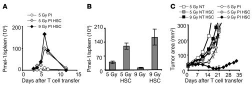
Figure 4. HSCs drive pmel-1 CD8+ T cell expansion in nonmyeloablated and myeloablated mice, but tumor treatment is only achieved in myeloablated mice.
(A and B) HSC transplantation–driven transgenic T cell expansion is not dependent on the intensity of immunodepletion. Mice received a preparative regimen of either 5 Gy or 9 Gy TBI, which was followed by effector pmel-1 CD8+ T cells (1 × 10⁶) and rhIL-2 with or without HSC transplantation. (A) Absolute numbers of adoptively transferred congenic marked pmel-1 CD8+ T cells in the spleen were enumerated on the days indicated. Results shown were derived from pooled splenocytes of 3 mice per group per time point (A) or from 3 individual mice (±SEM) assessed on day 6 (B) (P = 0.0009, 9 Gy versus 9 Gy/HSC; P = 0.02, 5 Gy versus 5 Gy/HSC; P = 0.24, 5 Gy/HSC versus 9 Gy/HSC). (C) HSC-driven expansion of pmel-1 CD8+ T cells is therapeutically effective in myeloablated, but not in nonmyeloablated, WT mice. Animals bearing tumors established for 10 days were either left as controls (NT) or received, prior to the transfer of 1 × 10⁶ effector pmel-1 CD8+ T cells and rhIL-2, either 5 Gy TBI, 5 Gy TBI and an HSC transplant, or 9 Gy TBI and an HSC transplant. Results for tumor area are the mean of measurements from 5 mice per group (±SEM). Results for each of the panels are representative of data from 3 independent experiments.

