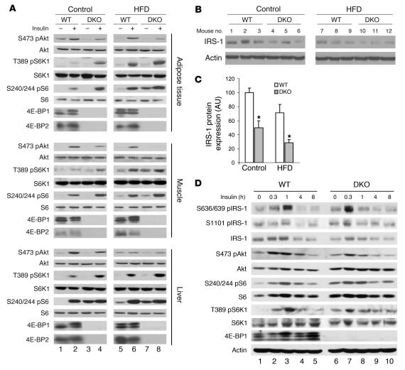
Figure 3. Deletion of 4E-BP1 and 4E-BP2 led to impairment of insulin signaling.
(A) Increased S6K activity and reduced Ser473 phosphorylation of Akt in muscle, liver, and adipose tissue from WT and DKO animals. Mice were fasted for 6 hours before receiving a 0.75 U/kg insulin injection in the tail vein. Animals were sacrificed 10 minutes later, and tissues were collected for Western blotting. An immunoblot of WT and DKO mouse tissue is shown. S473 pAkt, phosphorylated Ser473 of Akt; T389 pS6K1, phosphorylated Thr389 of S6K1; S240/244 pS6, phosphorylated Ser240/244 of S6. (B) Reduced IRS-1 expression in DKO adipose tissue. (C) Quantification of IRS-1 protein levels in WT and DKO adipose tissue. Levels were normalized to actin (n = 6–7). Data are mean ± SEM. *P < 0.05 versus WT (2-tailed, unpaired Student’s t test). (D) Immunoblot analysis showed increased inhibitory serine phosphorylation of IRS-1 (S636/639 pIRS-1 and S1101 pIRS-1) and sustained Thr389 phosphorylation of S6K1 in DKO MEFs following insulin treatment.

