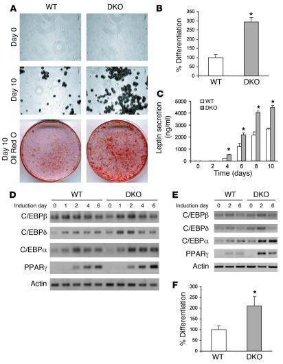
Figure 5. Deletion of 4E-BP1 and 4E-BP2 promotes adipocyte differentiation.
MEFs from WT and DKO embryos were grown to confluence and differentiated into adipocytes as described in Methods. (A) Microscopic images of MEFs following induction of adipogenesis. Original magnification, ×400. Lipid droplets were stained with oil red O solution. (B) Quantification of lipid incorporation by measurement of the intensity of oil red O staining in WT and DKO MEFs at day 10 of the adipocyte differentiation process. (C) Leptin secretion in the culture medium was measured throughout induction of adipogenesis and quantified. (D) RT-PCR analysis showing C/EBPβ, C/EBPδ, C/EBPα, and PPARγ expression following differentiation of WT and DKO MEFs. (E) RT-PCR analysis showing C/EBPβ, C/EBPδ, C/EBPα, and PPARγ expression following differentiation of preadipocytes isolated from WT and DKO adipose tissue. (F) Lipid incorporation was quantified by measuring the intensity of oil red O staining in WT and DKO preadipocytes at day 10 of the adipocyte differentiation process. Data are mean ± SEM from 4 different experiments. *P < 0.05 versus WT (2-tailed, unpaired Student’s t test).

