
Figure 5.
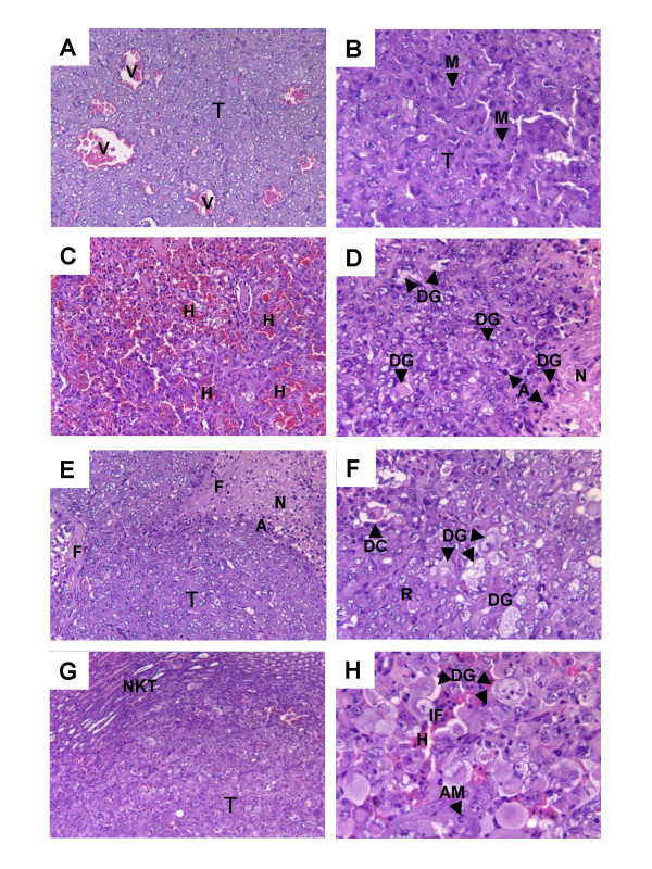
Histology of KCI-18 kidney tumors treated with genistein and radiation. Kidney tumors, resected from mice of the experiment described in Figure 4, were processed for histology and tumor sections were stained with H&E. The main findings were labeled on the prints with T for tumor, V for vessel, M for mitosis, H for hemorrhages, DG for degenerative, N for necrosis, A for apoptosis, F for fibrosis, DC for detached cells, R for rhabdoid cells, NKT for normal kidney tissue, IF for inflammatory cells and AM for abnormal mitosis. Panels A, B: Kidney tumor from control mice showing high-grade and very vascularized carcinoma (A, ×50) with frequent mitosis (B, ×100). Panels C, D: Kidney tumor from mice treated with genistein showing extensive hemorrhages (C, ×50), degenerative changes in tumor cells, apoptotic cells and areas of necrosis (D, ×100). Panels E, F: Irradiated kidney tumor, with areas of tumor destruction, showing fibrosis and apoptotic cells (E, ×50), focal areas with atypical detached rhabdoid cells (F, ×100). Panels G, H: Kidney tumor from mice treated with genistein and radiation, showing smaller residual tumor area adjacent to normal kidney tissue (G, ×25). The residual tumor looked hemorrhagic and consisted of large areas of detached rhabdoid cells, atypical giant cells with large nuclei and inflammatory cells (H, ×100). Lower and higher magnifications (×-fold) are presented to both show wider areas of tumor histology and focus on major findings.