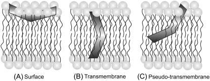
FIGURE 8.
A schematic representation of the membrane bilayer showing the various orientations and locations of membrane-bound melittin: (A) surface (2,29,48,68,70,72,74), (B) transmembrane (2,29,67,69,71,75), and (C) pseudo-transmembrane (50,51,73) orientations. The V-shaped bent rod represents the amphipathic, α-helical membrane-bound melittin. Our results using NBD fluorescence of NBD-melittin analogs support the surface (parallel) orientation (A) of membrane-bound melittin. Such a surface (parallel) orientation of melittin should be applicable to zwitterionic, anionic, and cholesterol-containing membranes under the experimental conditions used. See text for other details.

