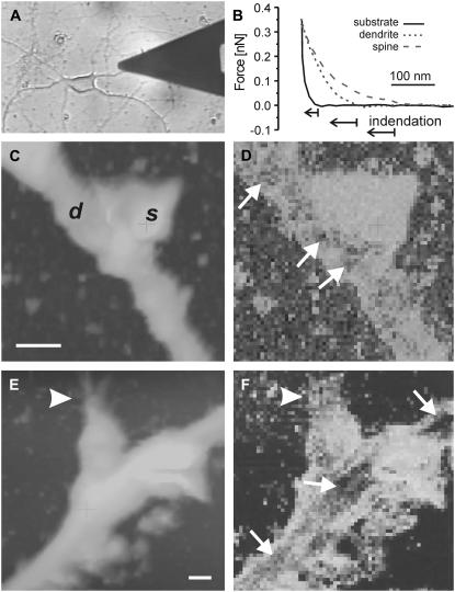
FIGURE 2.
Soft spines. (A) Photomicrograph of a cultured hippocampal neuron being scanned with an AFM probe. (B) Representative force-distance curves acquired during force-volume imaging of regions of a spine, dendrite, and substrate (as indicated). (C, E) Topography (under constant force) maps of two dendrites with spines (labeled d and s in C). Vertical color scale is 0.5 μm in C and 0.8 μm in E. Lateral bar is 1 μm in both. (D, F) Corresponding stiffness maps (bright is soft, dark is stiff). Spines appeared soft relative to the dendrite shafts, where stiff patches or fibers were identified (small arrows). Spine shapes were irregular, often exhibiting small surface protrusions (arrowheads). Axons were not observed in close proximity to the soft spines.

