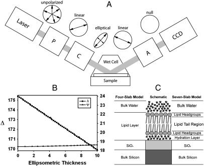
FIGURE 1.
Experimental setup forIE. (A) A schematic description of the PCSA (polarizer-compensator-sample-analyzer) reflection IE configuration used in this study. The windows of the wet cell are normal to the incident laser beam. The cartoon circles above each image illustrate the changes in polarization of the light under nulling conditions. (B) High sensitivity of Δ (and relative insensitivity of Ψ) on nanometer-scale thickness changes in the bilayer are revealed in a model calculation using a simplified four-slab model consisting of water/lipid phase/SiO2/Si. (C) A schematic of parallel-slab optical models for the SiO2/Si supported lipid bilayer (center) configuration sample systems considered in this study. A detailed seven-slab model (right) and a four-slab model approximation (left) used in our data analysis are also shown.

