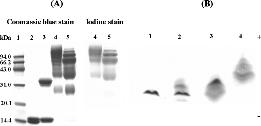
Figure 1. Characterization of PEGylated proteins by SDS/PAGE and IEF.
SDS/PAGE was carried out on a pre-cast 14% Tris/glycine gel from the Invitrogen Corporation. Lane 1, molecular mass markers; lane 2, HbA; lane 3, αα-fumaryl Hb; lane 4, (propyl-PEG5K)6-αα-Hb; lane 5, (propyl-PEG5K)6-Hb. Proteins were identified by Coomassie Blue staining, and PEG was detected by iodine staining. (Propyl-PEG5K)6-αα-Hb and (propyl-PEG5K)6-Hb were both loaded at the same amount of protein content (12 μg). IEF was operated using pre-cast resolve gels from Isolab and a blend of pH 6–8 resolve ampholytes. Lane 1, HbA; lane 2, αα-fumaryl Hb; lane 3, (propyl-PEG5K)6-Hb; lane 4, (propyl-PEG5K)6-αα-Hb.

