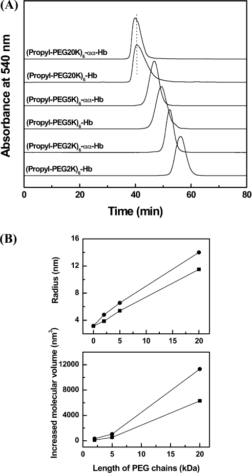
Figure 3. Influence of PEG chain length on the molecular volume of PEGylated αα-fumaryl Hbs.
(A) Size-exclusion chromatographic analysis of PEGylated protein. The analysis was carried out at room temperature on two HR10/30 Superose 12 columns connected in series. The columns were eluted with PBS, pH 7.4, at a flow rate of 0.5 ml/min. (B) Size enhancement of haemoglobin (■) and αα-fumaryl Hb (●) as a function of the length of attached PEG chains. The curves were made using Origin 6.0 software. Molecular radii were measured by dynamic light scattering at a protein concentration of 1 mg/ml. Increased molecular volume (ΔV) was calculated with an equation ΔV=4π(r3−r03)/3, where r and r0 are the radii of PEGylated haemoglobins and HbA respectively.

