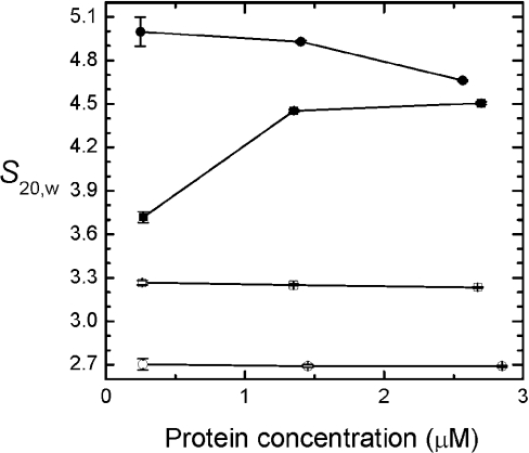
Figure 5. S20,W of PEGylated proteins as a function of haemoglobin concentration.
The curves were made using Origin 6.0 software. Sedimentation velocity measurements of (propyl-PEG5K)6-Hb (□), (propyl-PEG5K)6-αα-fumaryl Hb (○), HbA (■) and αα-fumaryl Hb (●) were conducted in a Beckman XL-I analytical ultracentrifuge in PBS, pH 7.4, at 25 °C and 55000 rev./min. Boundary movement was followed at 405 nm using the centrifuge's absorption optics.

