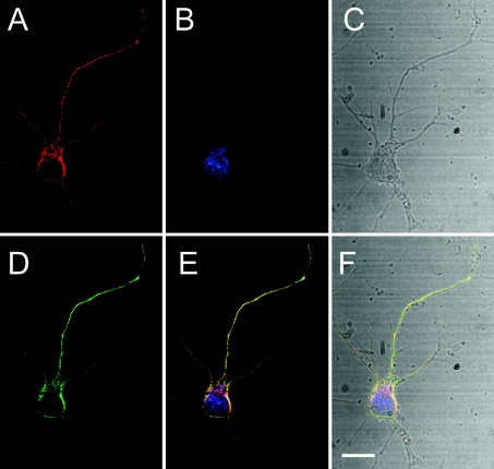
Figure 4. Ns partially co-localizes with chromogranin B.
Confocal microscopy of a primary mouse cortical neuron shows that the endogenous Ns in red (A) and chromogranin B in green (D) are in a neuron in blue (B). Transmission (grey) in (C) shows that this neuron is a single isolated cell. (E) The merged image of (A), (B) and (D). (F) The merged image of (A), (B), (C) and (D). Monoclonal anti-Ns antibody, polyclonal anti-chromogranin B antibodies and fluorescent Nissl stain were used to detect Ns, chromogranin B and neurons respectively. Secondary antibodies anti-rabbit Alexa Fluor® 488 and anti-mouse Alexa Fluor® 568 were used for fluorescence. All the images were deconvolved iteratively by using Volocity computer software. The scale bar shows 5 μm, and a ×100 oil-immersion objective lens was used.

