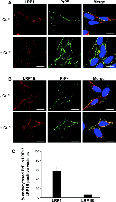
Figure 3. LRP1 but not LRP1B co-localizes with PrPC after its Cu2+-mediated endocytosis.
SH-SY5Y cells expressing PrPC were seeded on to glass coverslips and grown to 50% confluence. Cells were then pre-incubated with antibody 3F4 at a dilution of 1:1000 in PBS for 30 min at 4 °C, washed three times in PBS and then incubated for 20 min at 37 °C in OptiMEM in either the absence or the presence of 100 μM Cu2+. Cells were fixed, permeabilized and then incubated with either (A) a goat polyclonal antibody against LRP1 or (B) a rabbit polyclonal antibody against LRP1B overnight at 4 °C. Cells were then incubated with AlexaFluor® 488 conjugated rabbit anti-mouse antibody and then with either AlexaFluor® 594 conjugated rabbit anti-goat (for LRP1) or AlexaFluor® 594 conjugated goat anti-rabbit (for LRP1B). Nuclei were stained using DAPI (4′,6-diamidino-2-phenylindole). Cells were viewed using a DeltaVision Optical Restoration Microscopy system. Images are representative of three individual experiments. Scale bar=10 μm. (C) The co-localization of intracellular PrPC, internalized in response to Cu2+, with LRP1 or LRP1B was quantified using Imaris 4. The data represent the percentage of intracellular PrPC-containing vesicles that also stained positive for LRP1 or LRP1B (±S.E.M.); n≥10 cells.

