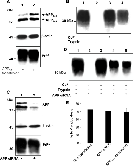
Figure 4. Cu2+-stimulated endocytosis of PrPC is unaltered either by overexpression or knockdown of APP.
(A) SH-SY5Y cells expressing PrPC were stably transfected with a pIREShyg vector containing the cDNA encoding APP751. Mock transfectants were created by transfection of an empty pIREShyg vector. Lysates were immunoblotted for APP, PrPC and β-actin. (B) Cells stably expressing APP751 were surface biotinylated and incubated with or without 100 μM Cu2+ for 20 min at 37 °C. Prior to lysis, the cells were incubated with trypsin to digest cell-surface PrPC. Cells were then lysed and total PrPC immunoprecipitated from the sample using antibody 3F4 and then subjected to Western blot analysis. The biotin-labelled PrPC fraction was detected with peroxidase-conjugated streptavidin. (C) SH-SY5Y cells expressing PrPC were incubated for 48 h with a 2 μM solution of the siRNA against APP, prepared with DharmaFECT-1 transfection reagent in OptiMEM. Mock-transfectants were incubated in the presence of DharmaFECT-1 only. Lysates were immunoblotted for APP, PrPC and β-actin. (D) Endocytosis experiment as described in (B) using the cells incubated in the presence or absence of APP siRNA. (E) Densitometric analysis (means±S.E.M.) for multiple blots from three separate experiments performed in (B) and (D).

