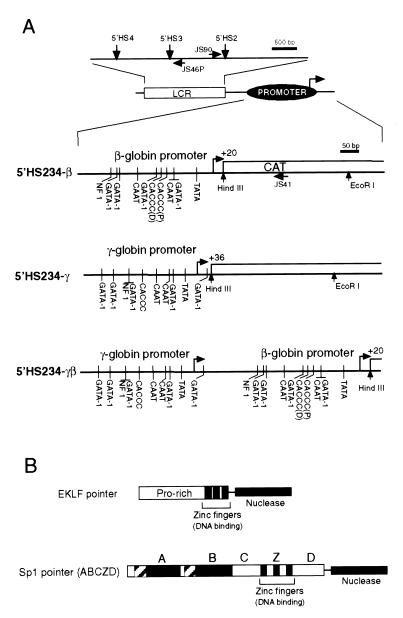
Figure 1.
(A) Diagram of the target plasmids. Downward arrows mark the positions of 5′HS2, 5′HS3, and 5′HS4 of the β-globin LCR (mini-LAR, mini-locus activating region), which is linked upstream of the β-globin (5′HS234-β), γ-globin, (5′HS234-γ), or tandem γ- and β- promoter. Horizontal arrows mark the positions of primers used for primer extension in this report, and vertical lines mark the positions of identified transcription factor binding sites in the β-globin promoter. Transcription initiation sites of both promoters are indicated with bent arrows. (B) Structure of EKLF and Sp1 pointers. The 25-kDa nuclease domain (black rectangle) of Fok I restriction endonuclease was fused to the carboxyl terminus of EKLF and Sp1. The positions of zinc fingers for EKLF and Sp1 (Z) are shown, as are the domains previously characterized for EKLF (Pro-rich, proline-rich) and Sp1 (domains A, B, C, and D).

