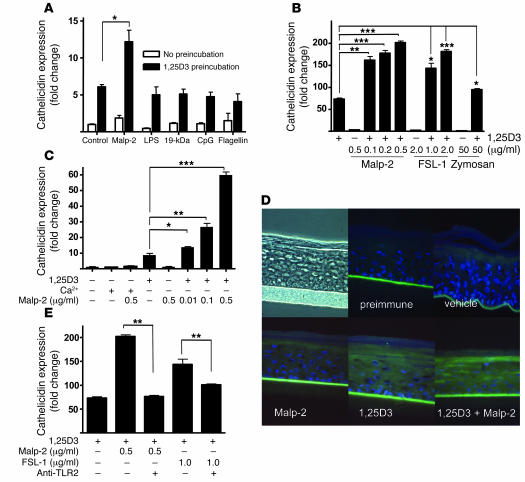
Figure 6. TLR2 function in keratinocytes is increased by 1,25D3.
(A) To test the functional relevance of the increased expression of TLR2 and CD14 in keratinocytes exposed to 1,25D3, cells were incubated with a low dose of 1,25D3 (0.1 nM for 24 hours) and then stimulated with TLR ligands for an additional 24 hours. Cathelicidin mRNA abundance was measured as in Figure 2. Only TLR2/6 ligand Malp-2 increased expression above that induced by low-dose 1,25D3 alone. (B) Dose-dependent response of cathelicidin expression following administration of TLR2/6 agonists Malp-2, FSL-1, or zymosan at the indicated concentrations in the presence of 1,25D3 (100 nM). (C) Cathelicidin mRNA expression in keratinocytes incubated with a low dose of 1,25D3 (0.1 nM) or a high calcium concentration (1.7 mM) for 24 hours and then stimulated with Malp-2 for another 24 hours. (D) Organotypic epidermal constructs were stimulated with Malp-2 (0.1 μg/ml) alone or in the presence of 1,25D3 (100 nM), and cathelicidin peptide expression was determined by immunostaining. Constructs were stained with a polyclonal anti–LL-37 antibody (green), and nuclei were detected with DAPI (blue). Original magnification, ×400. (E) Cathelicidin mRNA expression was inhibited by a TLR2-neutralizing antibody applied to keratinocytes stimulated with Malp-2 or FSL-1 in the presence of 1,25D3 (100 nM). Data are mean ± SD of a single experiment performed in triplicate and are representative of 3 independent experiments. *P < 0.05, **P < 0.01, ***P < 0.001, Student’s t test.

