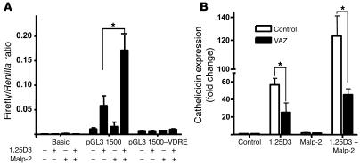
Figure 7. TLR2 activation signals cathelicidin induction through the vitamin D3 pathway.
(A) A functional VDRE was required for transcriptional activation of cathelicidin by Malp-2 and 1,25D3. HaCaT keratinocytes containing cathelicidin promoter reporter constructs (pGL3 1,500) were treated with Malp-2 (0.1 μg/ml) in the presence or absence of 1,25D3 (100 nM). Promoter constructs with a deleted VDRE at position –619 bp to –633 bp (pGL3 1,500–VDRE) lost transcriptional activity in all experiments. Values represent the ratio between firefly and Renilla luciferase activities. (B) In addition, treatment of keratinocytes with the VDR antagonist ZK159222 (10–7 M) blocked Malp-2–induced cathelicidin. Data are mean ± SD of a single experiment performed in triplicate and are representative of 3 independent experiments. *P < 0.05, Student’s t test.

