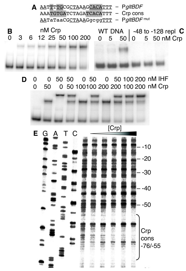
Figure 5.

Interactions between Crp and the gltBDF promoter region. A. The glt Crp-binding sequence (top) compared to the consensus Crp binding sequence (middle). The most-conserved elements of the Crp sequence are shaded, and matches to the consensus are underlined. The bottom sequence shows the mutations (lowercase) that were introduced for the experiment shown in Fig. 6. B. Gel mobility shift analysis of Crp binding to glt DNA region -406 to +246. The running buffer and gel contained 20 μM cAMP. C. Gel mobility shift analysis as in (B), except that two DNAs are used. One is WT, and the other has the region from -128 to -48 replaced by an equal length of exogenous DNA (amplified from the coding region of the cat gene). D. Gel mobility shift assay showing simultaneous binding of Crp and IHF to gltBDF DNA. A fragment of gltB DNA from -203 to +8 was used in the assay. E. DNAse I protection of glt DNA by Crp binding. A gltB DNA fragment from -203 to +161 labeled at the -203 end of the partner strand was used for the assay. The first 4 lanes represent the sequencing ladder of the partner strand. Crp concentrations of 0, 12.5, 25, 50, 100 and 200 nM were used in the assay.
