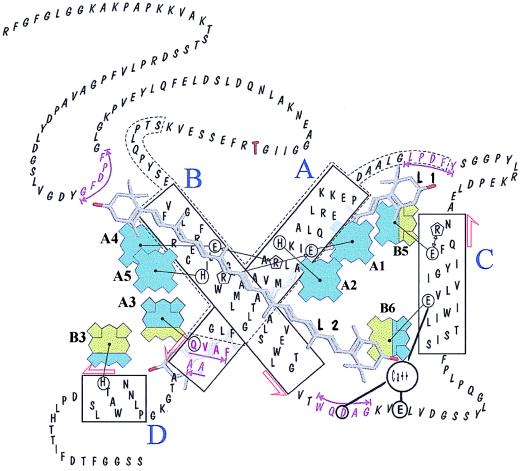
Figure 2.
Molecular model of the CP29 protein obtained by homology with LHCII (5) and on the basis of mutation analysis. Tetrapyrroles are shown in dark gray for Chl-a and in light gray for Chl-b. Sites that can be occupied by either Chl-a or Chl-b have mixed filling. The portions of helices A and B showing inner homology are contoured by a broken line. Putative xanthophyll-binding sequences are underlined, and the phosphorylation site is in bold. Black lines connect chemical groups that are thought to closely interact in the folded protein.

