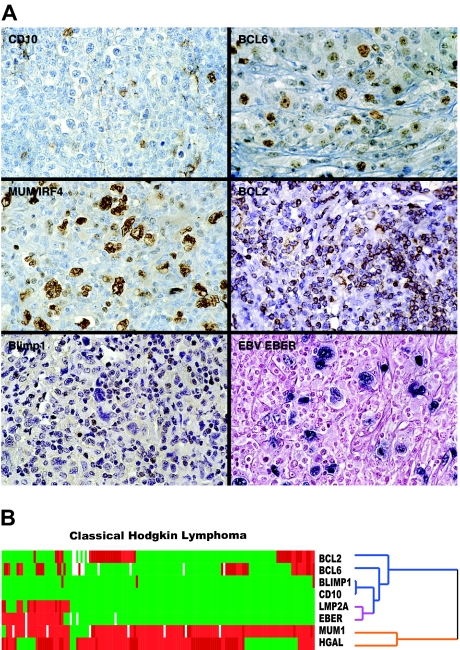
Figure 2.
Representative immunostains and EBV expression in classic Hodgkin lymphoma. (A) Typical examples of TMA cores of cHL stained for BCL6, CD10, BCL2, MUM1/IRF4, and Blimp1 and in situ hybridization for EBV EBER RNA are shown. (B) Hierarchic cluster analysis of immunohistologic and in situ hybridization data shows that HGAL clusters with MUM1/IRF4 on 1 branch of the dendrogram (orange) but not with BCL2, CD10, and BCL6 (blue) or with the 2 EBV-specific markers, LMP2A and EBER (purple), which cocluster with each other.

