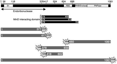
Fig. 1.
Schematic representation of RNaseE and Yfp-labeled RNaseE constructs. RNase domains are depicted as described in ref. 41. S1 domain (S1 RNA-binding domain), RBD (arginine rich RNA-binding domain), RhlB (RhlB-binding domain), enolase (enolase-binding domain), and PNPase (PNPase-binding domain) are shown. The region that includes the endoribonuclease catalytic domain is indicated (26). The black rectangles represent the RNaseE fragments that interacted with MinD in the yeast two-hybrid screen. The Yfp-labeled RNaseE constructs are shown in gray.

