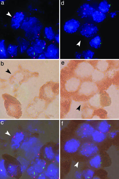
Fig. 2.
Female cells in male pancreas tissues. Immunohistochemistry for insulin and CD45 was used with concomitant FISH for X and Y chromosomes for the same cells as recently described (16). a, b, and c are from a boy with T1D and ketoacidosis; d, e, and f are from a boy with acute myeloblastic leukemia. (a) Fluorescence microscopy showing a female cell (arrowhead) with two (red) X chromosome signals. (Magnification: ×100.) Other cells contain one red and one Y chromosome (green) signal. Nuclei are identified with DAPI (blue). (b) Light microscopy of the same cells as in a. (Magnification: ×100.) The red-brown substrate identifies β cell insulin expression. (c) Overlay of a and b showing the identical cells with FISH and immunohistochemistry. (d) Fluorescence microscopy showing a female cell (arrowhead). (Magnification: ×100.) (e) Light microscopy of the same cells as in d. (Magnification: ×100.) (f) Overlay of d and e. Female cells were morphologically similar to surrounding β cells, and the cells did not express CD45.

