
Figure 3.
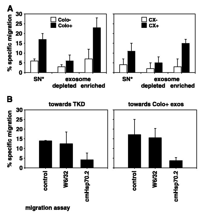
CD94+ NK cells specifically migrate toward Hsp70/Bag-4–positive exosomes derived from Colo+ and CX+ carcinoma sublines. A, VivaSpin treated (2-fold concentrated) supernatants (SN*) collected after a 24-hour cultivation period in serum-free medium, derived from Colo−/Colo+ (left) and CX−/CX+ (right) carcinoma sublines were used as positive controls for the migratory capacity of CD94+ NK cells. Exosome-depleted and exosome-enriched fractions derived from the same supernatants by ultracentrifugation at 150,000 × g were also used as attractants for NK cells. The transwell system consisted of two compartments separated by a polycarbonate membrane with a pore size of 5 μm. The different attractants, unseparated supernatant (SN*), exosome-depleted, and exosome-enriched fractions were placed in the lower compartment in a total volume of 600 μL; 0.5 × 10⁶ 51Cr-labeled CD94+ NK cells were placed in the upper compartment. After a 4-hour coincubation period at 37°C, the percentage of specifically migrating cells was determined in a γ-counter. B, migratory capacity of CD94+ NK cells was also tested toward Hsp70 peptide TKD (right) and exosomes derived from Colo+ carcinoma cells (left) either untreated or after preincubation with an MHC class I–specific (W6/32) or with an Hsp70-specific (cmHsp70.1) antibody. NK cells migrated specifically toward TKD and exosomes derived from Colo+ carcinoma cells. The MHC class I–specific antibody W6/32 did not affect migratory capacity; however, the Hsp70-specific antibody completely abrogated migratory capacity of NK cells toward TKD and Colo+ exosomes. Columns, mean of three independent experiments; bars, SE.