Figure 1. .
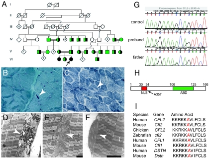
Pathologic and genetic findings in a family with CFL2 mutation A35T. A, Partial pedigree of the family illustrates several consanguineous loops. The proband is indicated by an arrow. The two affected sisters (filled circles) are homozygous for A35T, whereas an unaffected sister, both parents, and several other members of the extended family (half-filled circles) are heterozygous for the change and for a shared haplotype spanning ∼4.6-Mb pairs around the CFL2 gene. Green symbols indicate tested individuals with WT sequence. Light microscopic findings in proband’s muscle include presence of nemaline bodies (B, arrow) on Gomori trichrome staining and occasional minicores (C, arrow) on nicotinamide adenine nucleotide dehydrogenase-tetrazolium reductase staining. Electron microscopy confirmed identity of nemaline bodies (D, arrow), unstructured minicores (E, arrow), and concentric laminated bodies (F, arrow). G, DNA sequence analysis of genomic PCR products illustrating three genotypes for CFL2 c.103G→A seen in the family. H, Schematic representation of cofilin-2. Residue 35 is located next to NLS (30–34 aa); ABD = actin-binding domain. I, Altered alanine residue (red) is evolutionarily conserved among AC proteins (i.e., cofilin-2, cofilin-1, and destrin) across all sequenced vertebrates.

