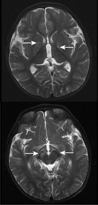
Figure 2. .
MRI scans made when patient 2 was 14 mo old (T2-weighted images). There is signal abnormality in the brain regions indicated by the white arrows: in the globi pallidi (upper panel) and in the midbrain with asymmetrical involvement of the cerebral peduncles (R→L) (lower panel). The appearances were considered likely to represent a neurometabolic disorder. No structural abnormality was noted.

