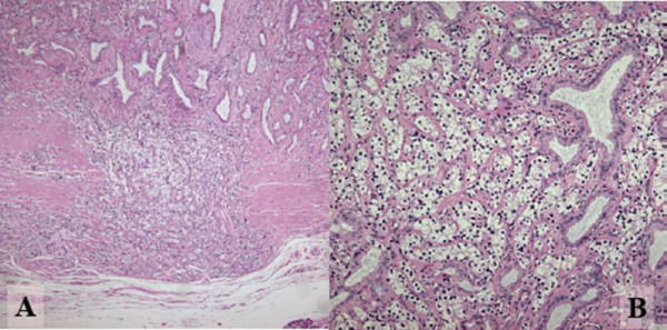
Figure 3.

Photomicrographs stained by Hematoxylin and Eosin. A: Tumor cells penetrating the fibromuscular layer of the bile duct, but not reaching to the surrounding pancreatic parenchyma. (Original magnification ×200). B: Tumor consisting of fairly uniform polygonal cells in size, with round nuclei and clear and abundant cytoplasm. Neoplastic cells are arranged in combination patterns with solid nests and trabecular growth. Preexisting non-neoplastic gland is entrapped in the lesion. (Original magnification ×400)
