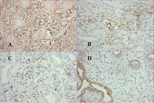
Figure 4.

Tumor cells were stained by Grimelius silver (A), and positive for immunohistochemical staining of Chromogranin-A (B) and for Synaptophysin (C). Clear cells were completely negative for Keratin, but positive in the intercalated non-neoplastic glands (D).
