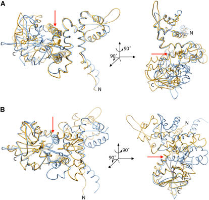
Figure 6.
Backbone Architecture of HI4′OMT.
(A) Cα alignments of the N-terminal portions of HI4′OMT (gold) and Ms I7OMT (blue) (residues 15 to 87 of both enzymes) result in an RMSD of 1.02 Å. Orientation of the right panel is achieved after a 90° rotation around an axis perpendicular to the plane of the page and a second 90° rotation around the vertical axis shown in the left panel as a red arrow.
(B) Cα alignments of the C-terminal portions (residues 330 to 364 of HI4′OMT and residues 318 to 352 of Ms I7OMT) result in an RMSD of 0.782 Å. Orientation of the right panel is achieved after a 90° rotation around an axis perpendicular to the plane of the page and a second 90° rotation around the vertical axis shown in the left panel as a red arrow.

