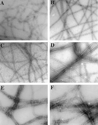
Figure 5.
Effect of SipA on the actin-bundling activity of T-plastin visualized by electron microscopy. Electron micrographs of negatively stained F-actin (2 μM) incubated with no (A and B), low (0.125 μM; C and D) or high (0.5 μM; E and F) concentrations of T-plastin in the presence (B, D, and F) or absence (A, C, and E) of SipA (2 μM). Highly organized F-actin bundles are visible in the presence of SipA and with low concentrations of T-plastin (C) which, by itself, does not induce actin-bundling. Tighter bundles are found in the actin bundles formed in the presence of SipA and high concentration of T-plastin (D), although the same concentration of SipA alone (B) had no effect on bundling. (Bar = 0.1 μm.)

