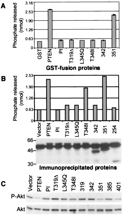
Figure 5.
The phosphatase activity of the PTEN C-terminal mutants. Shown is a phosphatase assay using 2.5 μg glutathione S-transferase-fusion proteins (A) or immunoprecipitated proteins from lysates of 293T cells transfected with FLAG-tagged PTEN and mutants (B). (B Lower) The amount of the immunoprecipitated proteins that, at the end of the phosphatase reaction, were resolved on SDS/PAGE and analyzed by immunoblotting with M2 antibody. The amount of free phosphate released in the reaction was measured in a colorimetric assay and compared with a standard curve. (C) PKB/Akt phosphorylation in U87-MG cells stably expressing PTEN and the indicated mutants. Proteins (50 μg) from total lysates were analyzed by immunoblotting with anti-PKB/Akt antibodies recognizing total levels of PKB/Akt (Akt) or only the phosphorylated form (P-Akt). These experiments demonstrated repeatedly similar results.

