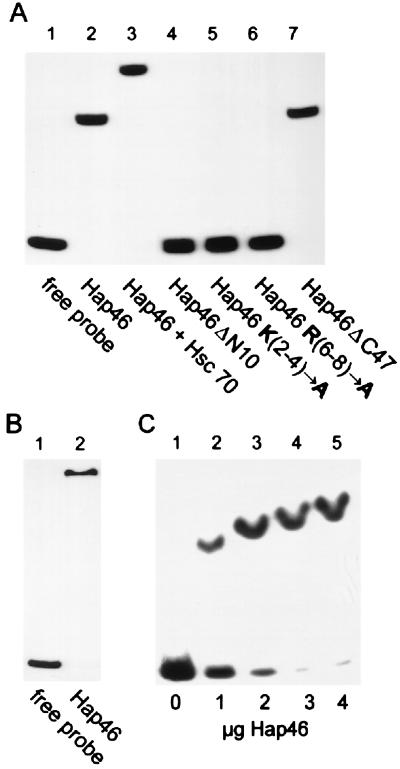
Figure 3.
Hap46 binding to DNA. (A) A radiolabeled 283-bp fragment of pcDNA3/CAT was either employed as such (lane 1) or used for electrophoretic mobility-shift assays with full-length Hap46 (lane 2), Hap46ΔN10 (lane 4), Hap46 K(2-4)→A (lane 5), Hap46 R(6-8)→A (lane 6), and Hap46ΔC47 (lane 7). In the experiment shown in lane 3, Hap46 was used in combination with hsc70. Analysis was by gel electrophoresis and autoradiography. (B) A 125-bp fragment from phage λ-DNA was subjected to the same assay with full-length Hap46 added (lane 2) or not (lane 1). (C) The 283-bp DNA fragment was used for gel-shift assays with either no protein added (lane 1) or with Hap46 at 1 μg (lane 2), 2 μg (lane 3), 3 μg (lane 4), or 4 μg (lane 5) per 30-μl assay.

