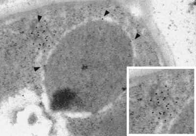
Figure 6.
NDC1p-3xpk is mislocalized when overexpressed. NDC1–3xpk was overexpressed by galactose induction, as described in Fig. 4. Immunoelectron microscopy analysis (see Materials and Methods) was used to examine the localization of overexpressed Ndc1p-3xpk. Shown is a cell at 40 min after release from the α-factor arrest in the presence of galactose. Arrows indicate membranous cytoplasmic structures and NPCs that display immunogold labeling, corresponding to Ndc1p-3xpk. Inset shows a closer view of the immunogold-labeled structure.

