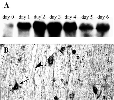
Figure 8.
Expression of Hic-5 in wild-type C2C12 cells. (A) Western blot analysis of endogenous Hic-5 protein levels in C2C12 myoblasts transferred to differentiation medium. Time is expressed as days in differentiation medium. (B) Immunohistochemical analysis of endogenous Hic-5 expression in C2C12 cells incubated in differentiation medium for 3 days. Expression is seen predominantly in mononucleated cells (arrow) as opposed to myotubes (arrowhead).

