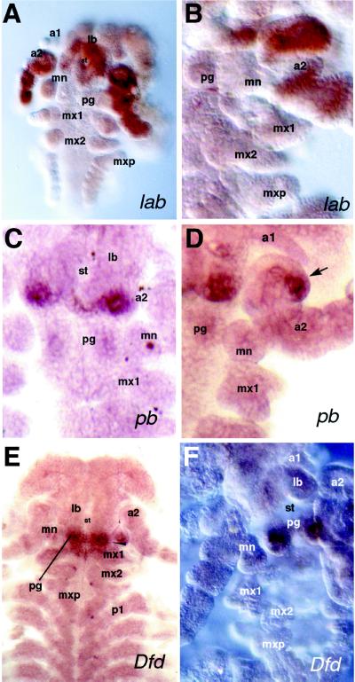
Figure 3.
Embryonic expression patterns of the P. scaber homologues of the Drosophila genes lab, pb, and Dfd, as revealed by in situ hybridization. Abbreviations as in Fig. 1. Ventral view of embryos with anterior at the top in all panels. (A and B) 50–60% Stage embryos showing the lab expression pattern; whole embryo (A) and close-up of embryonic head (B). (C and D) 50–60% Stage embryos showing pb expression in close-up views of the embryonic head revealing details of the expression domain. Arrow points to the pb expression in the posterior-lateral second antennae (D). (E and F) 50–60% Stage embryos showing Dfd mRNA distribution with dissected embryonic head (F) showing strong Dfd expression in the paragnaths. (A and E, ×50; B, C, D, and F, ×100.)

