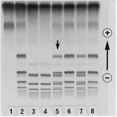
Figure 5.
A family study for the case II variant. HP–hemoglobin complexes were separated by a polyacrylamide gel and stained for peroxidase activity. The plasma samples were: lane 1, HP1-1; lane 2, HP2-1; lane 3, HP2-2; lane 4, mother; lane 5, propositus; lane 6, father; lane 7, mixture of equal amount of HP2-1 and HP2-2; and lane 8, HP2-1. Note that the pattern of the propositus (lane 5) was very similar to that of the mixture of HP2-1 and HP2-2 (lane 7).

