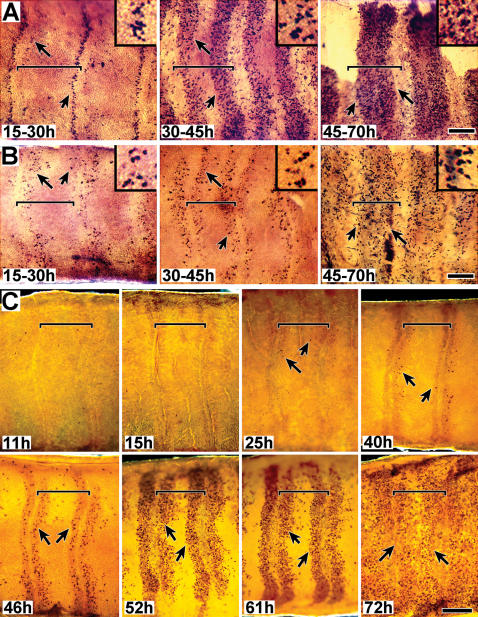
Figure 2. DNA replication in sensory epithelia. A,B. Mitotic activity studies using BrdU incorporation (male and female).
Developing sensory epithelia of male (A) and female (B) antennae are shown in whole-mount, attached to cuticle. Animals were injected with BrdU and tissue was dissected 15 hrs later; approximate injection and dissection times after pupation (a.p.) are indicated, based on developmental stages (stages 1–3). The tissue shown in 2B (45–72 h) is undergoing apolysis; a portion of the epithelium was torn during its removal and is thus missing. DNA replication was visualized using phosphatase tagged BrdU antiserum. Arrows indicate representative BrdU staining. Size bar (B, 45–70 h): 150 u (A–F); 50 u (inserts). C. Mitotic activity studies using phospho H3 visualization (male). Developing sensory epithelia, all male, are shown in whole-mount, attached to cuticle. Animals were dissected at the indicated times a.p. Nuclei undergoing mitosis were visualized using phospho-H3 antiserum. Arrows point to representative positive staining; no detection was observed prior to 24 hrs. Vertical lines visible along annular borders, especially noticeable in the 11h and 15h tissues, are part of the underlying (outer) pupal cuticle; annular borders align with these cuticular structural features. Size bar (C, 72 h): 200 u. For all figures (A–C), distal is left, proximal is to the right. Sensory epithelium was examined still attached to the outward facing antennal pupal cuticle. Annular boundaries and the proximal-distal orientation of the tissue were easily identified by distinct markings in the overlying pupal cuticle. Tissues were photographed in darkfield. Horizontal brackets mark the limits of single annuli within each panel; proximal is to the left and distal to the right.

