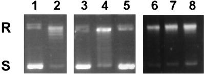
Figure 8.
Topoisomerase I and p53 are physically associated in mitomycin C-treated cells. MCF-7 breast carcinoma cells (wt p53) and HT-29 colon carcinoma cells (mutant p53) were treated with the DNA-damaging agent mitomycin C (10 μg/ml) for 4 hours followed by a chase in drug-free media for an additional 20 hours. Nuclear extracts were treated with PAb 1801 monoclonal anti-p53 antibodies, and the topoisomerase I activity associated with the p53 immunoprecipitates was determined by relaxation of supercoiled DNA. Lanes 3–8 show the catalytic activity of p53 immunoprecipitates from untreated control cells (lanes 3 and 6), cells treated with mitomycin C for 4 hours (lanes 4 and 7), or cells treated with mitomycin C for 4 hours followed by a 20-hour chase in drug-free media (lanes 5 and 8) obtained from either MCF-7 cells (lanes 3–5) or HT-29 cells (lanes 6–8). In comparison, the first two lanes show the migration of the DNA substrate alone (lane 1) or in the presence of purified topoisomerase I (lane 2). S, supercoiled DNA; R, relaxed DNA.

