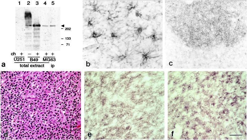
Figure 1.
(a) mAb 9.2.27 and rabbit 553 anti-rat NG2 antibodies recognize the same NG2 core protein. Extracts from human U251MG, MG63 and rat B49 cells lines before (−) and after (+) chondroitinase ABC (ch) treatment were subjected to Western blot by using the rabbit 553 anti-rat NG2 antibody. Lane 5 shows immunoprecipitates from MG63 cells that were formed with mAb 9.2.27. (b) mAb 9.2.27 recognizes complex process-bearing cells in human cerebral cortex resected for intractable epilepsy. (c–f) OLIGO express NG2 and PDGFα-R. At low magnification, NG2 immunostaining (c) demarcates a hypercellular neoplastic area. High magnification of hematoxylin/eosin-stained tissue (d) demonstrates densely packed round cells typical of OLIGO. The majority of neoplastic cells show a membrane-labeling pattern with NG2 (e) and PDGFα-R+ (f) antibodies. Tumor 30. [Bar = 35 μm (b), 120 μm (c), and 50 μm (d–f).]

