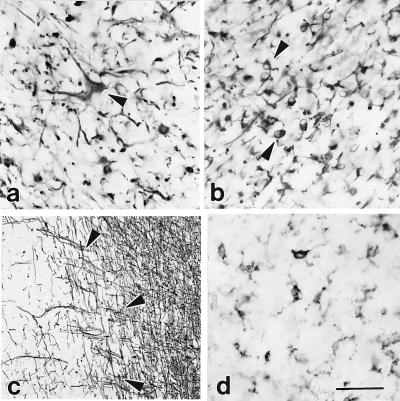
Figure 3.
OLIGO contain reactive astrocytes, neoplastic GFAP+ cells, residual myelin, and activated microglia. GFAP+ cells were present in all OLIGO (a and b). Many had the appearance of reactive astrocytes (a, arrowhead). Occasional cells had one or two short GFAP+ processes (b, arrowheads) and may reflect neoplastic cells. (c) MBP antibodies stained myelin in normal brain adjacent to OLIGO (Right) and residual myelin within the tumor (Left), but not neoplastic cells. Arrowheads denote the border between tumor and adjacent normal brain. (d) LCA+-activated microglia were seen frequently within the neoplasms. (a and b) Tumor 3. (c and d) Tumor 30. [Bar = 70 μm (a and b), 100 μm (c), and 60 μm (d).]

