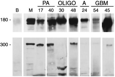
Figure 4.
Western blots detect PDGFα-R and NG2 in gliomas. (Upper) Although barely visible in normal brain tissue, PDGFα-R was detectable in extracts from all tumors examined. (Lower) A 300-kDa band corresponding to the NG2 core glycoprotein was present in half of the tumors. Lower-molecular-weight bands also were seen (see text). B, normal brain; M, human MG63 osteosarcoma cells.

