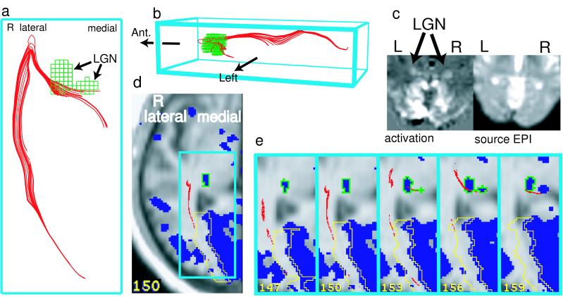
Figure 3.
Functional selection of geniculo-calcarine diffusion tracks by using fMRI activations (different subject from Fig. 1; Aσ ≥ 0.11). 3D projection views (a and b) of tracks (red) and fMRI-defined LGN filtering volumes (green) viewed directly from below (a) and from the left (b). In c, the visual cortex and LGN were activated by visual stimulation (unthresholded fMRI subtraction image is displayed above the corresponding source echo-planar image). Functional selection of tracks (d and e) used activated LGN and visual cortex regions. Tracks were selected anteriorly based on passage into the LGN volume (green outline), which was traced from the total activation thresholded at ≥0.22% signal change (blue region). Tracks were selected posteriorly in the occipital lobe based on passage into a border region (yellow), which was constructed as a 1-cm band lateral to the activation in medial occipital cortex (thresholded at ≥0.41%). Border filtration was implemented because of the absence of spatial overlap between tracks and visual cortex activation (where tracks terminated at white matter borders and activations were confined to gray matter).

