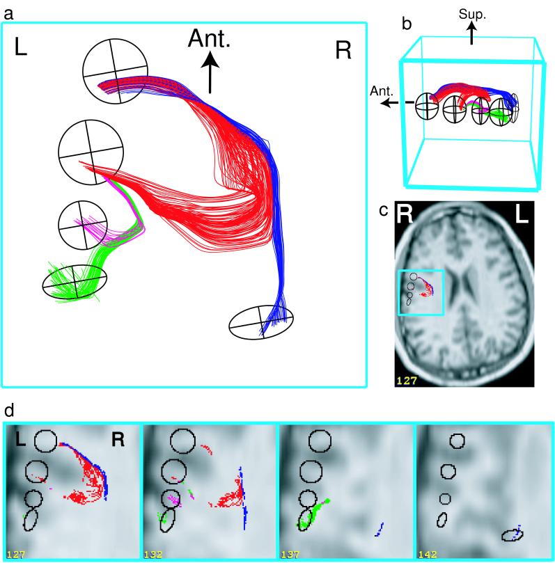
Figure 4.
Diffusion tracking of parietal association fibers (same subject as Fig. 1; Aσ ≥ 0.11). 3D projections (a and b) are viewed directly from above (a) and from the left-superior direction (b). In c, a 2D anatomical overlay onto one whole brain slice (#127) demonstrates general anatomical location. Magnified 2D overlays (d) demonstrate the fine anatomical location of tracks and filtering ellipsoids. Tracts were anatomically selected as those that entered paired combinations of five ellipsoids (black), four located in subcortical white matter, and one positioned in deep white matter. Of the 10 possible paired filtering combinations, the top four combinations (displayed) yielded 93% of the tracks (107 red, 44 green, 31 blue, and 15 magenta tracks). Three of the combinations yielded zero tracks. The blue tracks end in a region of below-threshold anisotropy in deep white matter.

