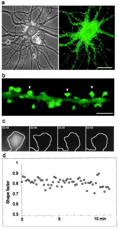
Figure 1.
Assessment of actin-dependent dendritic spine motility in living neurons. (a) Primary rat hippocampal neurons expressing GFP-tagged γ-cytoplasmic actin by transfection develop normally [phase contrast of a living cell (Left)] and acquire numerous dendritic spines after 3 wk in culture (Right). Bar = 30 μm. (b) GFP-actin targets to spine heads (arrowheads) where it is present at higher concentrations than in the dendrite shaft. Bar = 2.5 μm. (c) Images of a single dendritic spines in individual frames from time-lapse recording were processed by using a computer algorithm to produce profile outlines [original image and derived profile shown (Left)]. Selected profiles, taken 10 sec apart, demonstrate changes in spine shape that occur during recording. (d) Changes in the shape of single spines during time-lapse recordings were followed by calculating a shape factor from the spine profiles shown in c and plotting them against time, as shown in this example. A perfect circle has a shape factor of one, whereas values for flat objects approach zero.

