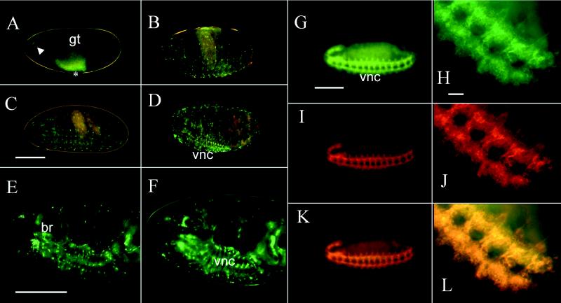
Figure 1.
GFP fluorescence and double-labeled antibody staining in embryos. A live stage-12 embryo (A) shows green fluorescent neurons in the anterior end (arrowhead) and in the ventral nerve cord (asterisk). The ventral nerve cord neurons are partially obscured by the strong yellow autofluorescence of the gut at this stage. In stage-15 embryos (B), some of the peripheral sensory neurons near the surface as well as many cells in the ventral nerve cord are visible. By stage 16 (C) and 17 (D) more cells in the ventral nerve cord and brain and a few peripheral sensory neurons show fluorescence. The same field of view is shown for a stage-17 embryo in E near the surface and F more internally. All embryos are oriented with anterior to the left and ventral to the bottom. A–D are color charge-coupled device images, E and F are false-colored black and white charge-coupled device images. Fixed, stage-12 embryos were double stained with anti-GFP (G and H) and anti-HRP (I and J) antibodies. The hazy green area in G is yellow autofluorescence of the gut. The complete nervous system is stained with anti-GFP antibody (G) and is shown at higher power (H) or is stained with anti-HRP antibody (I) and is shown at higher power (J). Images in G and I as well as H and J were merged to produce K and L, respectively. There is a complete correspondence of the anti-GFP and anti-HRP staining. Note also the difference between GFP fluorescence in a live stage-12 embryo (A) and anti-GFP antibody staining of a similarly aged fixed embryo (G and H). The images in G–L are false-colored. br, brain; gt, developing gut; vnc, ventral nerve cord. (Bar = 100 μm for whole embryos, 10 μm for higher power views in H, J, and L.)

