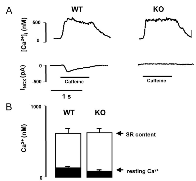
Figure 1.

Comparison of caffeine-induced Ca2+ transients and forward NCX currents in WT vs KO myocytes. A, [Ca2+]i transients and membrane current (INCX) recordings during caffeine application in patch clamped WT (n=8) and KO (n=8) myocytes loaded with fura-2 via the patch pipette. Cells were held at −40 mV and exposed to 5 mmol/L caffeine for 1 sec, using a rapid solution exchanger, to release SR Ca2+ stores. In WT, the Ca2+ release elicited an inward NCX current, which was absent in 8 of 9 KO myocytes. B, Summary graph showing that resting [Ca2+]i and the peak of the [Ca2+]i transient, and therefore SR Ca2+ load, were similar in both groups.
