
Figure 5.
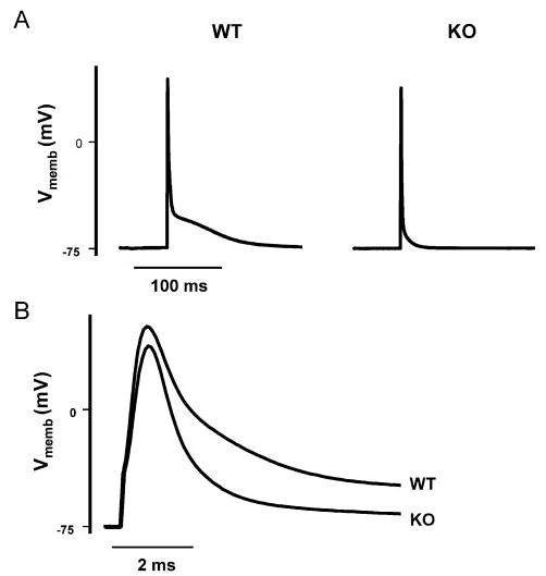
Faster AP repolarization in KO vs WT myocytes. AP tracings are representative for 9 KO and 10 WT myocytes. The pipette solution contained (in mmol/L): 110 KCl, 5 MgCl, 10 NaCl, and 20 HEPES (pH 7.2) with KOH. A, KO APs lack a plateau phase as compared with WT. B, First 10 ms of the tracings shown in A displayed on an expanded time scale. Vmemb indicates membrane potential.